The Kidney Biopsy – The Role in Diagnosis and Clinical Investigation
The evolution of kidney biopsies culture techniques was instrumental in the advancement of nephrology, providing unprecedented understanding of kidney anatomy; biochemistry and pathology [1]. The biopsy sample can be obtained by percutaneous or open biopsy during surgery, usually with guidance from an ultrasound for the former and visual identification for the latter. Regardless of the inherent, albeit low risks, kidney biopsies provide valuable information for the diagnosis of complex pathologies as well as monitoring transplant rejection [2-4]. A cross-section of the kidney renal cortex and a micrograph of a tissue slice can be seen in Figure 1.
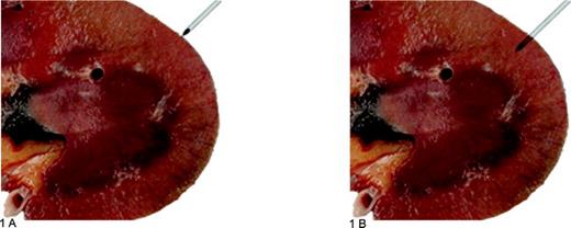
Figure 1: The cross-sectional anatomy of the kidney (a) shows the correct placement of a biopsy needle, where the correct needle size needs to be used to obtain the best biopsy sample (c) with volume and anatomical preservation. The renal cortex (b) shows red circular structures which denote intact of typical glomeruli. Taken and adapted from [2].
A kidney biopsy will be requested by the consulting physician when conventional methods such as urine and blood tests, ultrasound, X-rays as well as physical examination does not provide a definitive diagnosis or the results of tests indicate an underlying pathology which requires visual or microscopy confirmation. A biopsy provides information on [1-4]:
- Disease progression or treatment efficacy;
- The permanence of physical damage to the anatomical structures of the kidney by mechanical (e.g. crushing damage) or chemical injury (e.g. accidental ingestion of poison);
- Monitoring the effect of a therapy, new or existing, on the kidney;
- Monitoring the effect of chronic disease on the kidney (e.g. high blood pressure, obesity, diabetes, etc.)
- Diagnosing an acute or chronic disease of the kidney (e.g. cancer, fibrosis, etc.)
- Evaluation of transplant acceptance or rejection;
The use of kidney biopsies has advanced beyond clinical diagnosis and disease monitoring, receiving more critical use in advanced translational research aimed towards precision pharmacology and precision medicine. Biopsy-derived ex vivo models have proved crucial to advance investigation for absorption, distribution, metabolism, excretion and toxicity (ADMET) for drug development and drug discovery.
How to culture vascularized & immunocompetent 3D models in a standard Multiwell
Ex-vivo Kidney Biopsies Culture for Research and Precision Pharmacology
The use of kidney biopsy tissue as ex vivo disease models is a viable alternative to prolonged in vivo models, providing a sufficiently complex tissue microenvironment which can be readily observed in real-time. Using precision cut tissue slices (PCTS); 1-4mm in diameter and 200-400µm thickness; allows ex vivo culturing of renal tissues in static for up to 4 days without the loss of phenotype [5-9]. Approaches such as this allowed for researchers to investigate the molecular pathways of xenobiotics [5], drug metabolism [5, 6, 7] as well as acute and chronic kidney diseases [6, 7, 9-11]. Similarly, the use of precision cut tissue slices was demonstrated to enable a more precise efficacy screen to determine a therapeutic strategy for renal cancer patients [12].
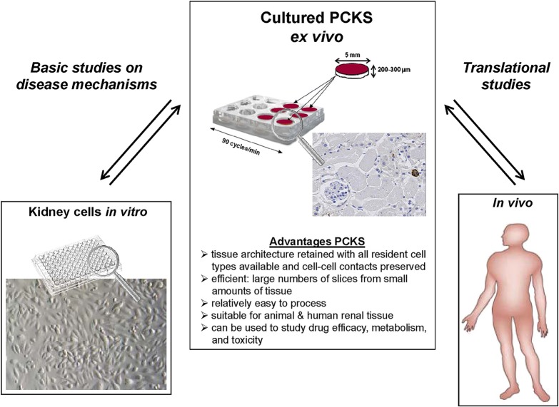
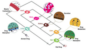
Figure 2: The advantages of precision cut tissue slices for kidney research. Taken and adapted from [6].
Precision cut kidney slices have been prepared using tissue from rodent [6-8] and human [9-12] origin, providing a genotype-phenotype correlation, relevant to the unique individual, in a cost-effective and timely conservative manner. Ex vivo culturing of biopsy tissues provide a complexity that is currently not achievable in other methods of 2D and 3D culturing, presenting with preserved cellular complexity and immune function [6]. Currently, efforts are ongoing to allow for the co-culture of precision cut slices from various organs in an organ-on-chip / biopsy-on-chip approach in a serial or parallel manner to more accurately recapitulate the complexity of drug metabolism [5]. However, the in vitro culturing of ex vivo derived precision cut slices requires methodology and technology standardization to allow for larger scale adoption in academia and translational researchers.
The Role of Oxygen Gradients in the Kidney
The kidneys are a metabolic demanding organ, requiring regular oxygen supply for the various metabolic and physiological functions performed. Additionally, oxygen exchange between the vascular and arterial systems occur in the kidneys which further promotes the need for oxygen homeostasis with regulation by hypoxia inducible factors (HIF) [13-15]. Kidney physiology require differential oxygen supply, where the proximal tubule cells are especially sensitive to hypoxia/ischemia-induced injury to the kidneys and are associated with acute and chronic kidney injury [13-16] as well as other pathologies such as fibrosis [13, 15, 16].
Two isoforms of HIF are present in the kidneys, where HIF-1Α is expressed for oxygen deprivation conditions less than 72h and HIF-2Α for prolonger oxygen starvation conditions. The HIF transcription factors regulate oxygen delivery, energy metabolism responses to oxygen as well as iron- and nitric oxide metabolism. Specifically, HIF-1Α regulates metabolism during glycolytic-oxidative metabolism switching for ATP synthesis; cytochrome C subunit conversion; inflammatory signalling and cell death signalling. HIF-2Α regulates the production of erythropoietin and implicated in angiogenesis [13, 14, 16]. The broad role of HIF in chronic kidney disease (CKD) can be seen in Figure 3.
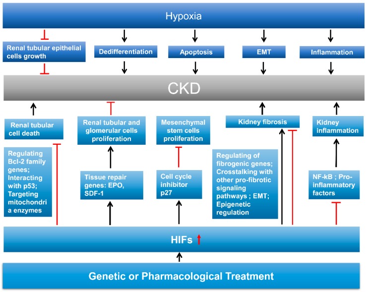
Figure 3: The interplay between hypoxia, HIF and chronic kidney diseases. Taken from [13].
Liu et al [17] recently demonstrated that HIF-1Α associated hypoxic events are correlated with progression of renal fibrosis through a p53-mediated pathway and cell cycle arrest in G2/M. The tumour suppressor gene p53 may be the link between acute or chronic kidney disease and kidney tumorigenesis, providing a preventative intervention therapeutic target for renal cancers.
References
- Cameron, J. S. and Hicks, M. J.The introduction of renal biopsy into nephrology from 1901 to 1961: a paradigm of the forming of nephrology by technology.Am. J. Nephrol., 17:347–358.
- Walker, P.D. The Renal Biopsy. Arch. Pathol. Lab. Med., 133:181-188.
- Shenbagaraman, P. and Connon, T. Kidney Biopsy Information for Patients. https://www.ouh.nhs.uk/patient-guide/leaflets/default.aspx
- Viscontim L. et al. Renal Biopsy : Still A Landmark for the Nephrologist. World. J. Nephrol., 5(4):321-327.
- De Kanter, R. et al. Precision-Cut Organ Slices as a Tool to Study Toxicity and Metabolism of Xenobiotics with Special Reference to Non-Hepatic Tissues. Current Drug Metabolism, 3:39-59.
- Poosti, F., et al. Precision-Cut Kidney Slices (PCKS) to study Development of Renal Fibrosis and Efficacy of Drug Targeting Ex Vivo. Disease Models & Mechanisms, 8:1227-1236.
- Genovese, F. et al. Precision-Cut Kidney Slices as a Tool to Understand the Dynamics of Extracellular Matrix Remodelling in Renal Fibrosis. Biomarker Insights, 11:77-84.
- Ihermann-Hella, A. and Kuure, S. Mouse Ex Vivo Kidney Culture Methods: Methods and Protocols. Methods in Molecular Biology(DOI:10.1007/978-1-4939-9021-4_2).
- Stribos, E.G.D. et al. Renal Fibrosis in Precision-Cut Kidney Slices. European Journal of Pharmacology, 790(5):57-61.
- Jensen, M.S. et al. Activation of the Prostaglandin E2EP2 Receptor Attenuates Renal Fibrosis in Unilateral Ureteral Obstructed Mice and Human Kidney Slices. Acta Physiologica, 227:e13291.
- Stribos, E.G.D., et al. Precision-Cut Human Kidney Slices as a Model to Elucidate the Process of Renal Fibrosis. Transl. Res., 170:8-16.
- Roelants, C. et al. Ex Vivo Treatment of Tumor Tissue Slices as a Predictive Preclinical Method to Evaluate Targeted Therapies for Patients with Renal Carcinoma. Cancers, 12:232.
- Shu, S. et al. Hypoxia and Hypoxia-Inducible Factors in Kidney Injury and Repair. Cells, 8:207.
- Haase, V.H. Mechanisms of Hypoxia Responses in Renal Tissue. J. Am. Soc. Nephrol., 24:537-541.
- Palm, F. et al. Uremia Induces Abnormal Oxygen Consumption in Tubules and Aggravates Chronic Hypoxia of the Kidney via Oxidative Stress. Am. J. Physiol. Renal. Physiol., 299:F380-F386.
- Rosenberger, C. et al. Hypoxia-Inducible Factors and Tubular Cell Survival in Isolated Perfused Kidneys. Kidney International, 70:60-70.
- Liu, L. et al. P53 Upregulated by HIF-1 Promotes Hypoxia-Induced G2/M Arrest and Renal Fibrosis In Vitro and In Vivo. Journal of Molecular Cell Biology, 11(5):371-382.

