Introduction
Spinning disk confocal microscopy is a particular configuration of confocal microscopy that is able to imaging at a faster rate. For this reason spinning disk confocal microscopy is particularly useful when imaging living samples or recording fast phenomena. Aim of this short scientific note is to introduce the technologies beneath spinning disk
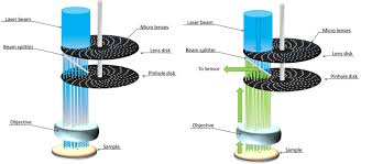
Fig 1, Optical path of light in spinning disk confocal microscopy. (Left) the laser beam passes trough a first Nipkov disks (Lens disk). Each hole has a microlens that concentrates the light at the level of a second disk. The holes in this second disk constitute the pinholes. Light with the right angle go to the sample passing trough the objective. (Right) Fluorescent light emitted by the sample is collected by the objective and filtered by the pinhole disk. A dichroic beam splitter placed between the two disks is then responsible in sending the sample image to the sensor.
How to culture vascularized & immunocompetent 3D models in a standard Multiwell
What is making spinning disk confocal microscopy faster than laser scanned confocal microscopy?
How spinning disk confocal microscopes make possible to observe fast physiological phenomena
Spinning disk confocal microscopy core technology consists of one or two Nipkov disks. In the most common two disk configuration the two disks have specific functions (Figure 2). The disk that in the optical scheme is closer to the sample has hundreds of pinholes and is used to illuminate and collect light from a specific focal plane. The second disk has also hundreds of small holes; each small holes host a micro lens that collects photons coming from the main laser beam. The photons are the collimated by the micro lenses in the pinholes of the first disk. An electrical motor accelerated the disks at high speed, this generate the specific illumination of spinning disk confocal microscopes.
Laser scanned confocal microscopy pushed significantly further the resolution in the three axes (x,y and especially z) when compared to standard fluorescence microscopy . Nevertheless laser scanner confocal microscopy drawback is the time needed to acquire good imaging since the laser spot produced by the pinhole has to scan the sample (Figure 2). Speed of the scanning is limited by the response time of the galvanic mirrors which speed is physically limited. Considering that quality of the image is inversely related to the speed of the scan and positively depending on the number of passages time to get good images can easily go up to one second per frame. The way laser scanner confocal microscopes illuminate the sample and collect the returning fluorescence implies the need of dedicated software to process the signals in order to reconstruct the image. On the other hand spinning disk confocal microscopes are significantly faster being able to obtain more than three hundred frames per second. This because sample is simultaneously illuminated by thousands of light spots (figure 2) and the returning signal can be directly collected by CCD sensors. By setting the acquisition speed of the CCD is possible to collect enough data to directly obtain the full focal plane image without any software algorithm. Another advantage of using a CCD sensor instead of a photomultiplier is that CCD sensors, although less sensitive, are much less keen to noise.
It has been empirically observed that spinning disk confocal microscopy has less photoxicity and is less keen to photobleach fluorophores when compared to laser scanner confocal microscopy. Photo damage or photo bleaching depend more on the intensity (dose/time) rather that the overall quantity of photons (absolute dose). The probability of secondary photochemical reactions is much higher in the presence of a short but strong photon flux (laser scanner confocal microscopy) whereas illuminating one spot several time with a lower amount of photons at time (Spinning disk confocal microscopy) results in less secondary reactions.
For those two reasons, which are faster and gentler image acquisition, spinning confocal microscopy is now considered the gold standard method to observe sample with a small to medium thickness.
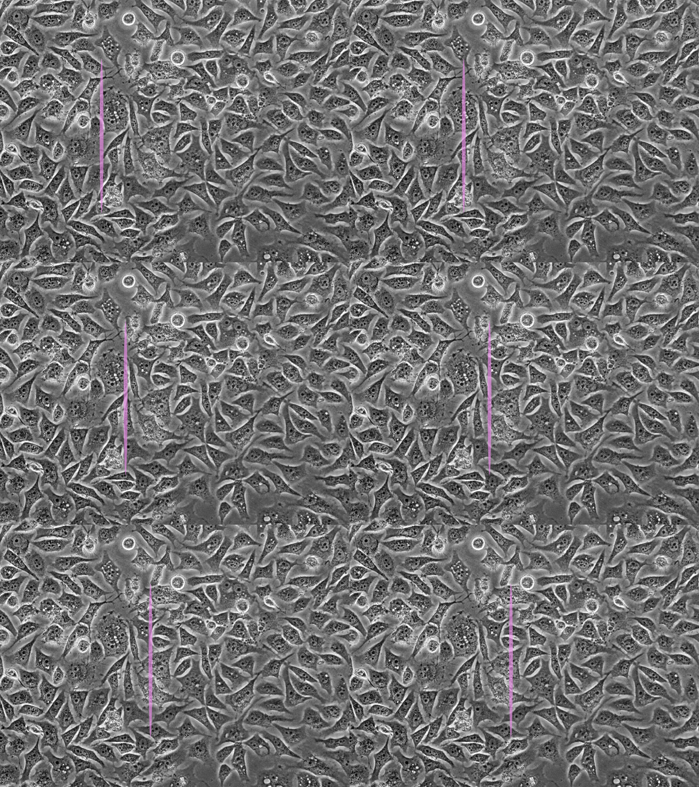
Figure 2. Animated simulation of laser scanning confocal microscopy (left) and spinning disk confocal microscopy (right) illumination. While in standard confocal microscopes the laser beam is manipulated in order to form a sort of line front scanning the sample, spinning disk confocal microscopes generate thousands of spots at time covering the whole sample area in a much shorter time.

