Introduction
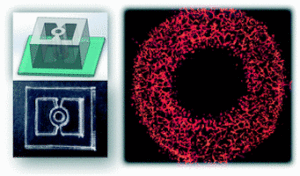
Human vascular microphysiological systems (MPS) represent promising three-dimensional in vitro models of normal and diseased vascular tissue. These systems, which mimic essential functional components of organs and tissues, draw on breakthroughs in tissue engineering, microfluidics, and stem cell differentiation.

Vascular models for microvasculature and medium-sized arterioles have already been established. Key vascular system functions have been replicated, and stem cells have the ability to mimic hereditary disorders and population diversity in genes that may raise an individual’s risk of cardiovascular disease. These systems can be used to assess novel treatment alternatives. Learn more in the paper featured below.
How to culture vascularized & immunocompetent 3D models in a standard Multiwell
Abstract
The authors state that “Microphysiological systems (MPSs), based on microfabrication technologies and cell culture, can faithfully recapitulate the complex physiology of various tissues. However, 3D tissues formed using MPS have limitations in size and accessibility; their use in regenerative medicine is, therefore, still challenging.
Here, an MPS-inspired scale-up vascularized engineered tissue construct that can be used in regenerative medicine is designed. Endothelial cell-laden hydrogels are sandwiched between two through-hole membranes. The microhole array in the through-hole membranes enables the molecular transport across the hydrogel layer, allowing long-term cell culture. 3D Microphysiological System Vascularized Tissue
Furthermore, the time-controlled delamination of through-hole membranes enables the harvesting of cell-cultured hydrogel constructs without damaging the capillary network. Importantly, when the tissue constructs are implanted in a mouse ischemic model, they protect against necrosis and promoted functional recovery to a greater extent than implanted cells, hydrogels, and simple gel–cell mixtures.”.
References
Bang, S., Tahk, D., Choi, Y. H., Lee, S., Lim, J., Lee, S., Kim, B., Kim, H. N., Hwang, N. S., & Jeon, N. L. (2021). 3D Microphysiological System‐Inspired Scalable Vascularized Tissue Constructs for Regenerative Medicine. Advanced Functional Materials, 2105475. https://doi.org/10.1002/adfm.202105475


