 Single-Cell Protein Atlas of Transcription Factors in C. elegans ...
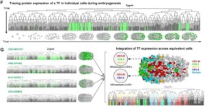
For the first time Zhuo Du et al. correlate mRNA and protein expression in C. elegans embryo to build the first embryo protein mapping. Afte...
Read more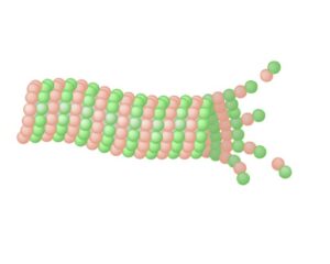 Microtubule Depolymerization By Cold...
Microtubules (MT) are involved in multiple cellular processes, including mitosis, cell transport, morphogenesis and motility. Two populatio...
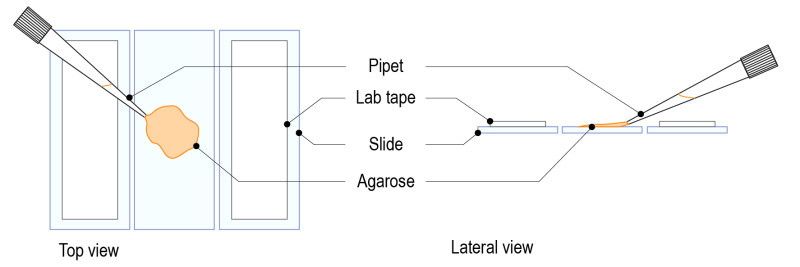
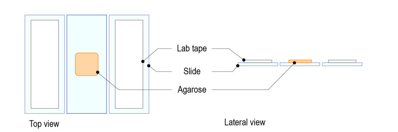
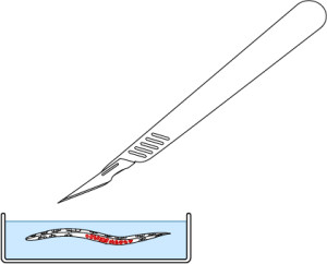
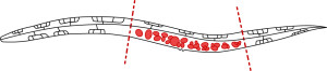
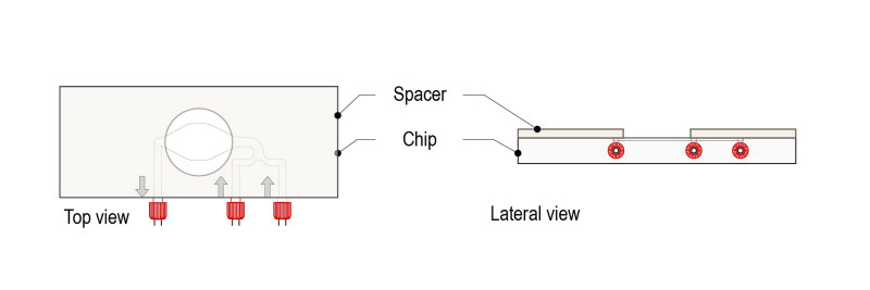
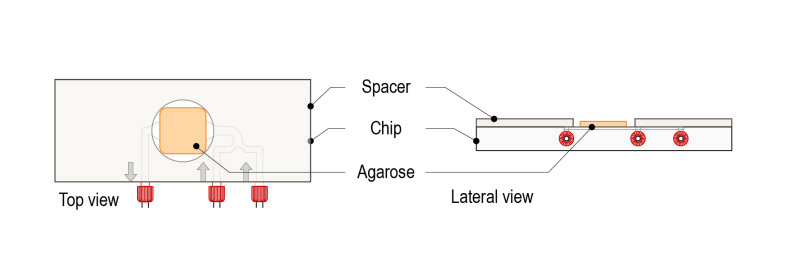
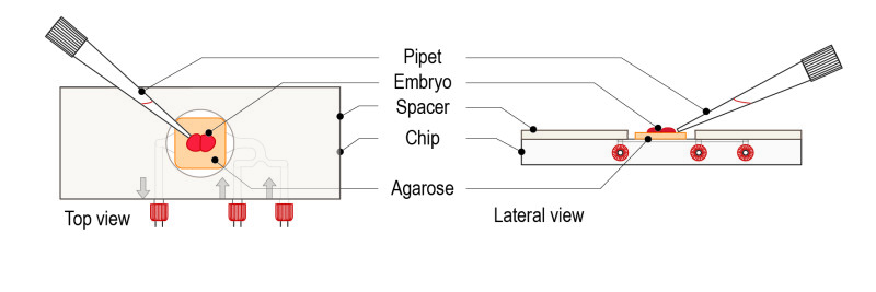
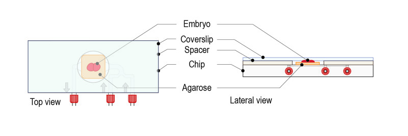
Read more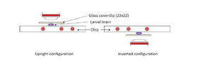 Hanging drop method for C. elegans embryos...
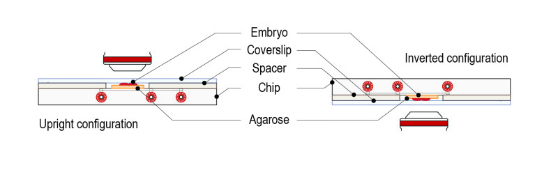
Introduction In this protocol, we show how to prepare C. elegans embryos on a standard coverslip to perform experiments with CherryTemp usin...
Read more