 Imaging Drosophila Third Instar Larval Brain Cells Expressing Jup...
Experimental conditions Drosophila brains expressing a centriole marker (GFP-PACT) and a microtubule marker (Jupiter-mCherry) were dissected...
Read more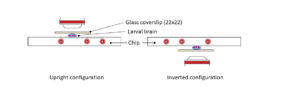 Protocol for Drosophila larval brains...
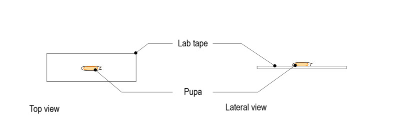
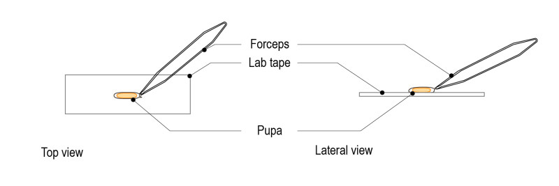
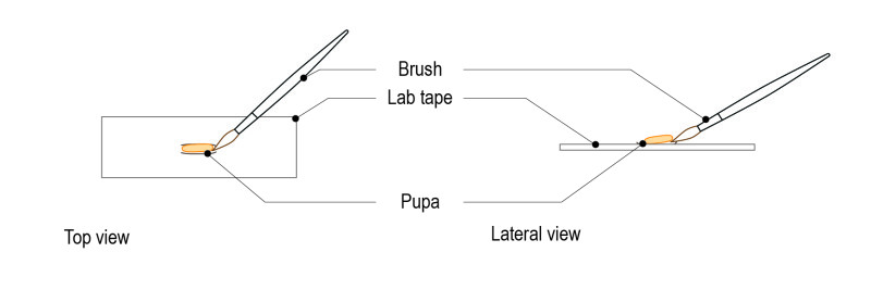
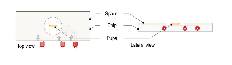
Mounting method for third larval brains of D. Melanogaster Introduction This protocol shows how to mount Drosophila 3d instar larval brains ...
Read more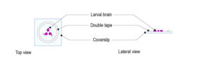 Protocol for Drosophila With Gas Permeable Chamber...
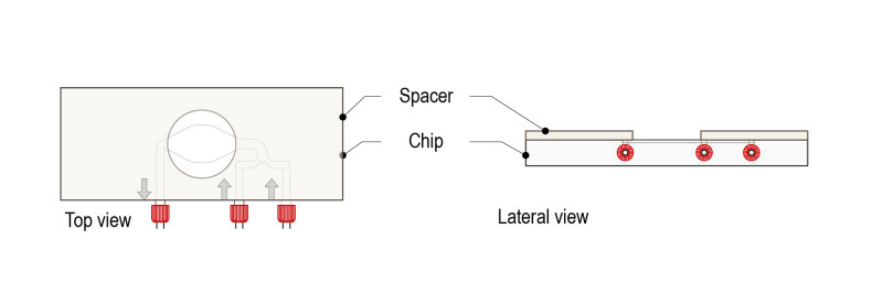
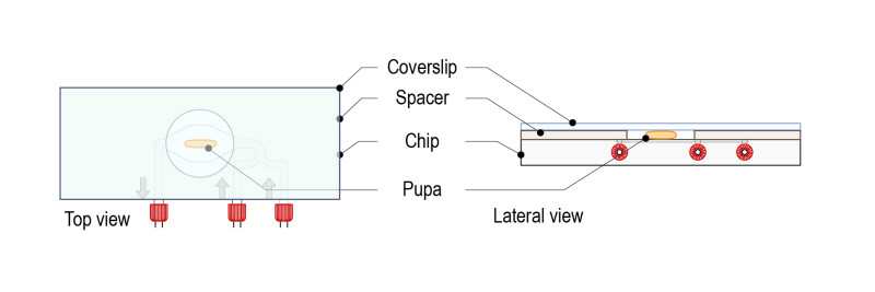
Mounting method for Drosophila larval brain Introduction With this protocol, a gas permeable chamber is created by means of a double-sided t...
Read more