Introduction
In this protocol, we show how to prepare C. elegans embryos on a standard coverslip to perform experiments with CherryTemp using the hanging drop method. It shows step by step how to dissect and mount the sample for the experiment.
Imaging Drosophila third instar larval brain cells expressing Jupiter-mCherry as a microtubule marker
Step-by-step guidelines:
Step 1: Pick young adults into pre-cooled M9 medium (16°C).

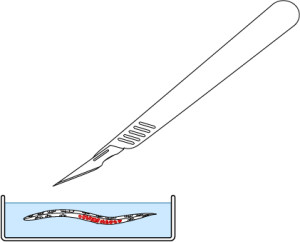
Step 2: Dissect them by cutting at both ends of the uterus using a scalpel and #15 blade to release the embryos.

Step 3:
Choose the embryos of the desired stage and transfer them to a cover slip using a mouth pipette.

Step 4:
Use a syringe to make a ring of Vaseline on a coverslip. Make sure to center the ring so that the embryos will be correctly placed under the thermalisation pattern of our chip.

Step 5:
Use a mouth pipette to transfer the embryos, cluster embryos with an eyelash tool.

Step 6:
Carefully cover with our temperature control chip. Gently clamp the system using our insert.

Step 7:
Place the mounted slide on either an upright or an inverted configuration by simply rotating 180°.

References
- Methods adapted from J. Dumont and J.C. Canman: https://www.sciencedirect.com/science/article/abs/pii/S0091679X16300619
- T. Davies, S. Sundaramoorthy, S.N. Jordan,M. Shirasu-Hiza, J. Dumont and J.C. Canman, 2016, “Using fast-acting temperature-sensitive mutants to study cell division in Caenorhabditis elegans“, Methods in Cell Biology, Volume 137, ISSN 0091-679X, http://dx.doi.org/10.1016/bs.mcb.2016.05.004


