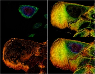
Wide-field fluorescence microscopy (Fluorescent microscopy)
Introduction to fluorescence microscopy Fluorescence is a natural phenomenon in which following the absorption of light

Introduction to fluorescence microscopy Fluorescence is a natural phenomenon in which following the absorption of light
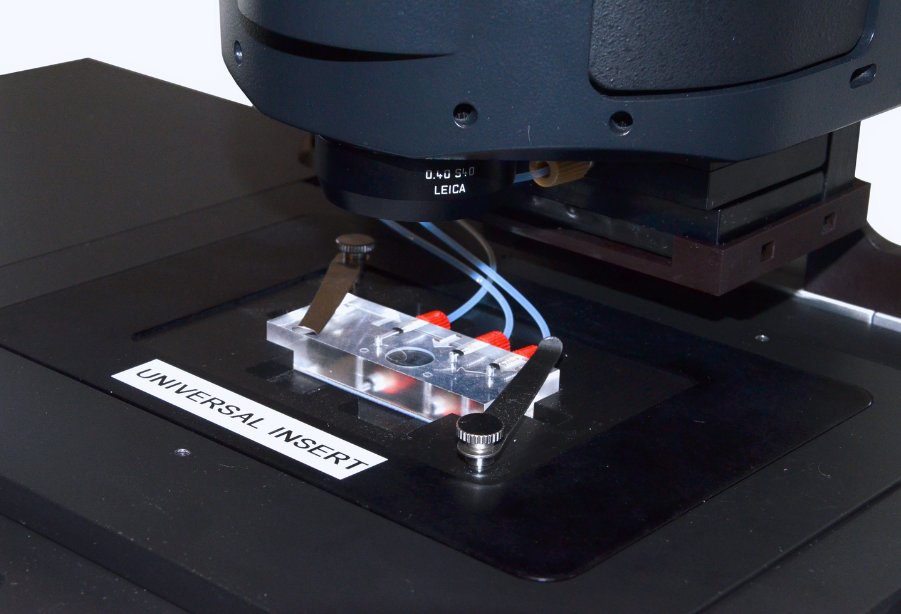
Experimental conditions Drosophila brains expressing a centriole marker (GFP-PACT) and a microtubule marker (Jupiter-mCherry) were dissected
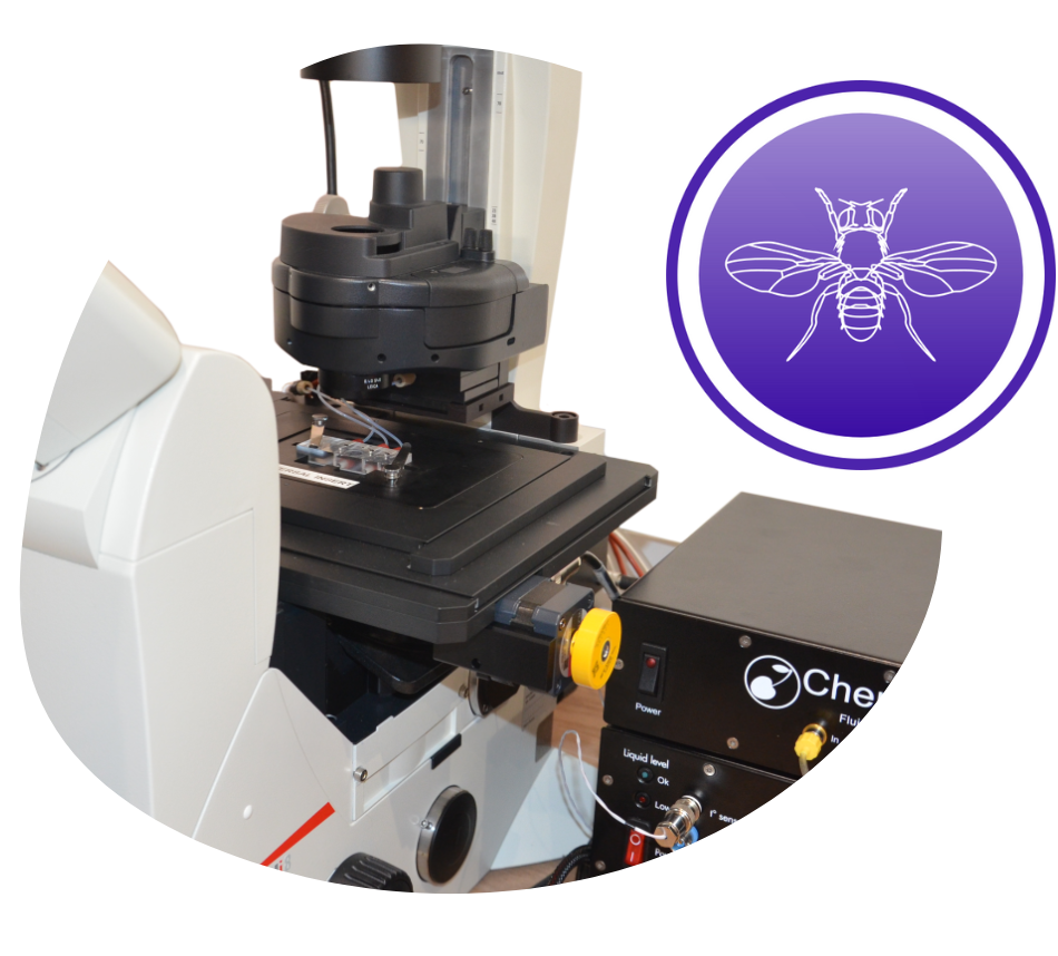
Experimental conditions Drosophila third instar larval brains expressing a microtubule marker (Jupiter-mCherry) were mounted on a
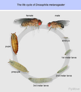
Introduction The use of D. melanogaster as a model organism in developmental biology has been an
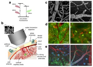
Introduction Two-photon excitation microscopy is a particularly microscopy technique based on the capability, under specific circumstances,
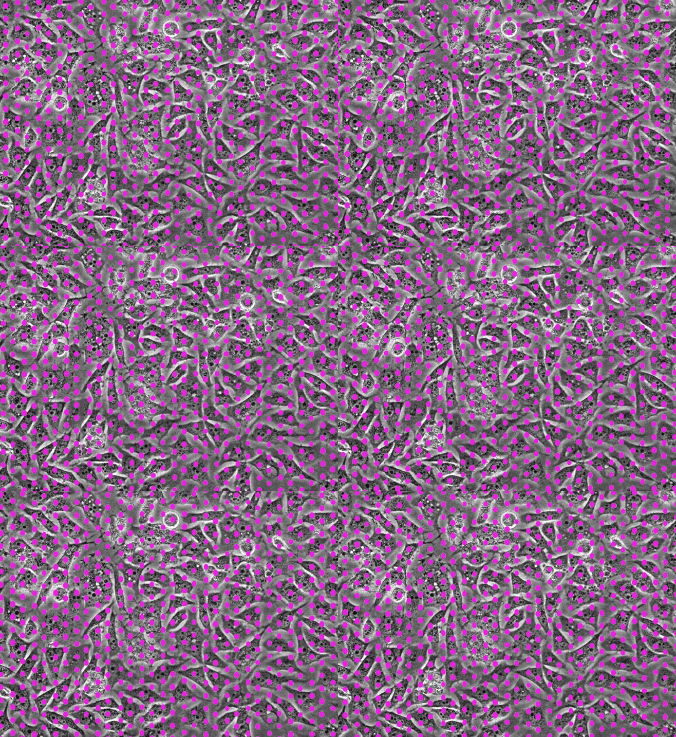
Introduction Spinning disk confocal microscopy is a particular configuration of confocal microscopy that is able to imaging at a
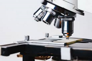
Introduction Non-adherent cells are difficult to image because of movement. This is the case of living
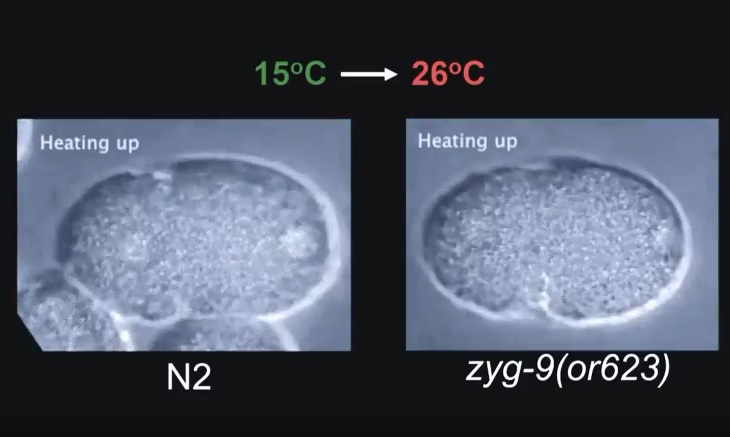
Experiment This video shows the temperature impact upon division of temperature-sensitive mutant C. elegans embryo (zyg-9).
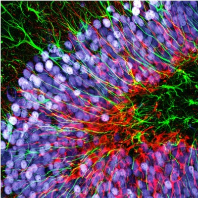
Introduction to confocal microscopy Laser scanning confocal microscopy, more generally referred as confocal microscopy, is an
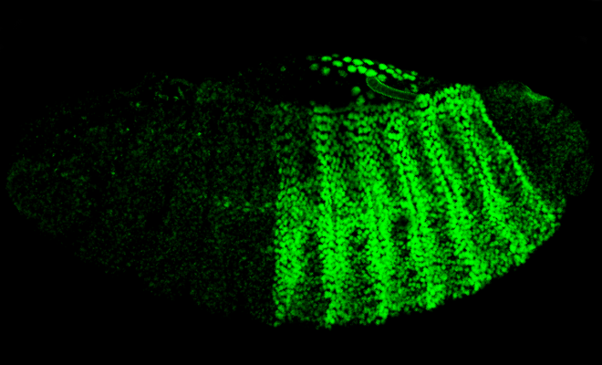
Introduction Drosophila embryo development or embryogenesis is a fast process during the life cycle of the
14 rue de la Beaune,
93100 Montreuil (Paris)
France
+33 9 87 04 70 35
contact@cherrybiotech.com